19 Mar Microfluidic Chemiluminescence Platform for Embryo Implantation Prediction
Assessing which embryos have the highest chance of successful implantation remains a major challenge in assisted reproduction. Current clinical practice relies heavily on morphological grading, which is subjective and often fails to accurately predict developmental potential. While metabolite analysis of embryo culture media offers a non-invasive alternative, conventional techniques such as mass spectrometry or HPLC are complex, require large instruments, and are not well suited for small sample volumes typical in IVF workflows.
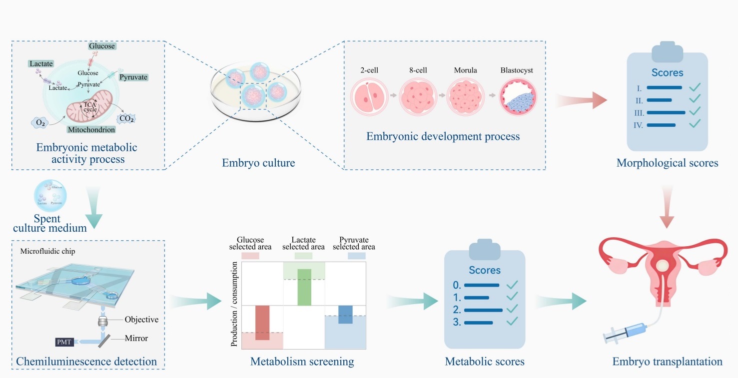
To address this gap, the authors developed a capillary-driven chemiluminescent microfluidic chip that enables simultaneous quantification of key embryo metabolites including glucose, lactate, and pyruvate. The microfluidic system integrates dielectric wetting valves that allow precise, on-demand fluid control without external pumps. By combining metabolic measurements with morphological data, they constructed a predictive model for embryo implantation potential with strong clinical performance.
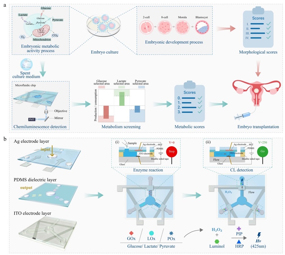
“Schematic of the metabolite detection process for embryo developmental potential assessment. (a) The morphology and metabolite comprehensive analysis based on a chemiluminescence dielectric valve chip. (b) The sample detection process. (i) The sample flowed to the enzyme reaction zone and halted before the valve, where it reacted with the preloaded enzyme. (ii) The dielectric valve was activated, allowing fluid to resume flow and triggering the chemiluminescence reaction.” Reproduced from Tong, W., Shi, J., Yu, Z. et al. Embryo metabolite analysis and implantation potential prediction using chemiluminescent microfluidic chips with dielectric wetting valves. Nat Commun (2026). under a Creative Commons Attribution 4.0 International License.
The microfluidic device is microfabricated as a multilayer microfluidic platform consisting of patterned electrodes, a PDMS dielectric layer, and microchannels formed using adhesive layers. Fluid flow is driven by capillary forces through the microfluidic device and halted at hydrophobic dielectric valves until a voltage is applied. Once activated, the microfluidic valves allow sequential flow into reaction zones where enzymes specific to each metabolite generate hydrogen peroxide, triggering chemiluminescence through luminol oxidation. This microfluidic design enables multiplexed detection within microliter-scale samples while maintaining high sensitivity and a wide detection range.
Operationally, only about 3 μL of spent blastocyst culture medium is required per assay. The microfluidic system achieves low limits of detection down to sub-micromolar levels and maintains linearity across a broad concentration range, accommodating the large differences between metabolites such as millimolar glucose and micromolar pyruvate. The chip also demonstrates good reproducibility, selectivity, and stability over time, making it suitable for practical use in clinical environments.
Using this microfluidic platform, the authors analyzed embryo metabolism in both training and validation cohorts. They found that embryos leading to successful pregnancies consumed more glucose and pyruvate while producing higher levels of lactate. These metabolic differences were statistically significant and consistent across datasets. Importantly, metabolic activity showed little correlation with traditional morphology scores, indicating that metabolite profiling provides complementary information rather than redundant insight.
Building on these findings, the team developed a multi-parameter predictive model that integrates metabolic and morphological data. The metabolite-based model alone achieved strong predictive performance, and when combined with morphology, the overall model reached an area under the curve (AUC) of 92.0%. This significantly outperformed morphology-only approaches, demonstrating the added value of incorporating metabolic information into embryo selection workflows.
In conclusion, this study presents a compact and sensitive microfluidic platform for non-invasive embryo assessment. By enabling rapid and multiplexed metabolite analysis from minimal sample volumes, the system provides a practical route toward improving embryo selection in IVF. With further validation in larger cohorts, this approach could support more informed clinical decision-making and improve implantation success rates.
Figures are reproduced from Tong, W., Shi, J., Yu, Z. et al. Embryo metabolite analysis and implantation potential prediction using chemiluminescent microfluidic chips with dielectric wetting valves. Nat Commun (2026). https://doi.org/10.1038/s41467-026-69999-5 under a Creative Commons Attribution 4.0 International License.
Read the original article: Embryo metabolite analysis and implantation potential prediction using chemiluminescent microfluidic chips with dielectric wetting valves
For more insights into the world of microfluidics and its burgeoning applications in biomedical research, stay tuned to our blog and explore the limitless possibilities that this technology unfolds. If you need high quality microfluidics chip for your experiments, do not hesitate to contact us.