24 Aug Microfluidic nano-plasmonic imaging platform for purification- and label-free single small extracellular vesicle characterization
The detection and analysis of small extracellular vesicles (sEVs), such as exosomes, has attracted significant attention due to their potential as circulating biomarkers for diseases, especially cancers and liver disorders. Yet, their small size and the complexity of blood plasma make direct detection difficult. As the authors note,
“conventional sEV characterization methods… typically require prior isolation and purification, which are time-consuming, yield low recovery rates, and lack reproducibility, thus making their translation toward clinical applications challenging.”
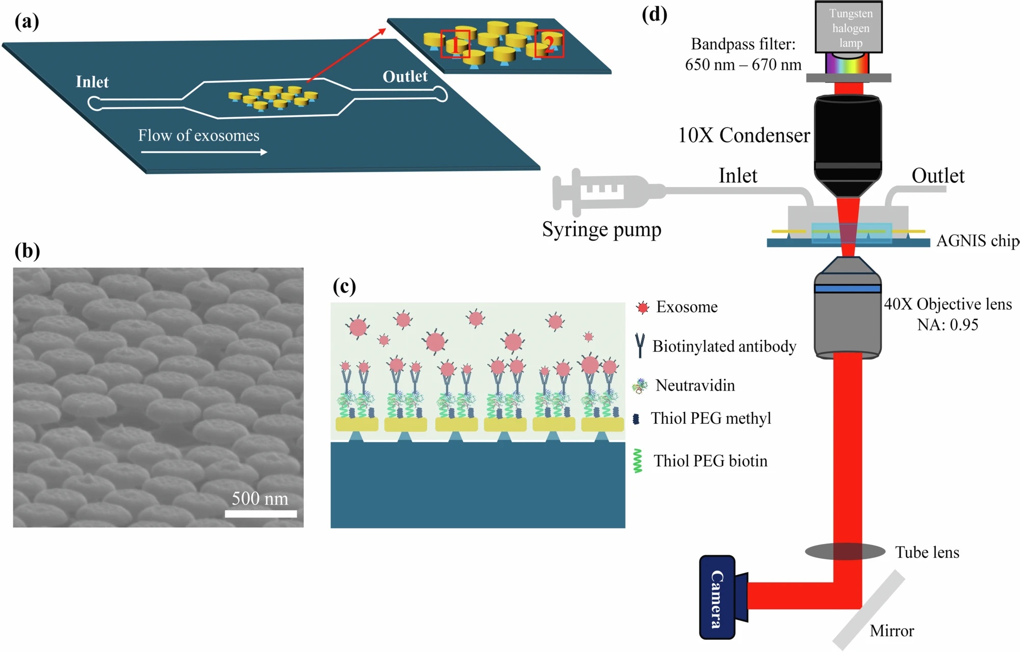
To address this challenge, researchers present a microfluidic chip integrated with PlAsmonic NanO-apeRture lAbel-free iMAging (PANORAMA). This platform enables direct, purification-free, label-free characterization of single sEVs in plasma. The microfluidic device combines automated fluid handling with a plasmonic sensing surface made of arrayed gold nanodisks on invisible substrates (AGNIS), functionalized with antibodies (CD63, CD9, CD81) that selectively capture vesicles. The microfluidic design also incorporates a “push–pull” flow strategy to increase interactions between vesicles and the sensing surface, improving sensitivity and reproducibility.
The team microfabricated the microfluidic AGNIS surface and functionalized it with exosome-specific antibodies. By flowing plasma or purified vesicle samples through the microfluidic channel under controlled push–pull conditions, they captured vesicles directly on the plasmonic substrate. PANORAMA imaging detects binding events by monitoring local refractive index changes as vesicles adhere, translating into optical contrast signals at the single-vesicle level.
“a Conceptual illustration of the microfluidic device. b Scanning electron microscope (SEM) image of the AGNIS surface. c Schematic of AGNIS surface functionalization for exosome capture from human plasma. d Schematic of the optical setup for PANORAMA with the microfluidic device at the sample position.” Reproduced from Mohsen Daraei, O., Singh, A.K., Mohapatra, S. et al. Microfluidic nano-plasmonic imaging platform for purification- and label-free single small extracellular vesicle characterization. npj Biosensing 2, 26 (2025). under Attribution 4.0 International License.
Their results demonstrated precise and reproducible detection of vesicles. Using purified exosome samples, they recorded time-lapse images over 60 minutes, with particle counts rising steadily to over 500 detections within a sensing area, while maintaining low background noise. When applied directly to patient plasma without purification, the system detected and quantified hundreds of sEVs within an hour, with accurate size estimation (~130–140 nm) consistent with vesicle biology. Importantly, the platform required only 20 μL of plasma and completed analysis within 60 minutes, highlighting its efficiency compared to conventional techniques.
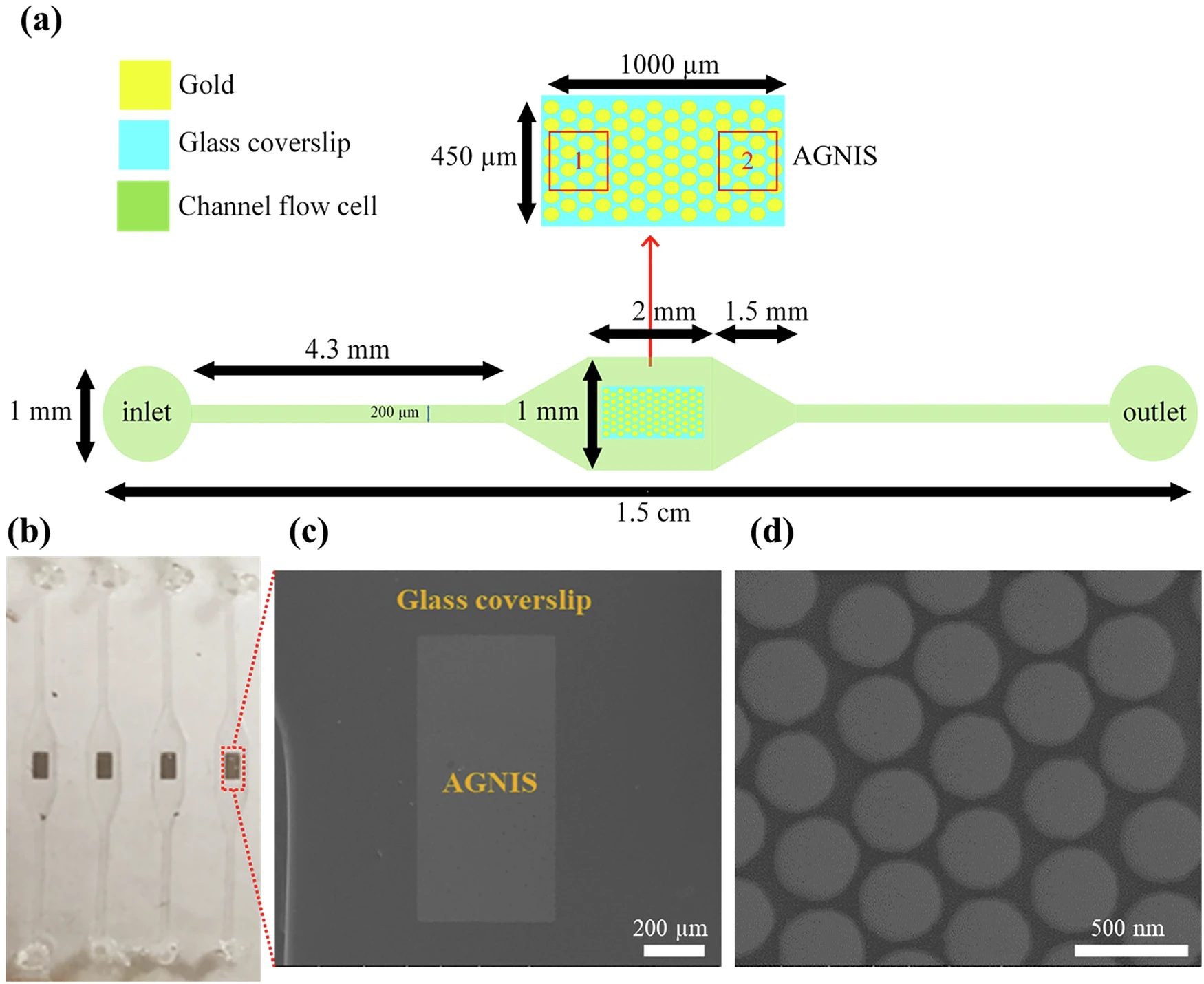
“a Microfluidic channel design. b Optical image of the AGNIS-integrated microfluidic device. c SEM image of a single AGNIS patch within the microfluidic channel. d SEM image (top view) of densely packed AGNIS from a small area in (c).” Reproduced from Mohsen Daraei, O., Singh, A.K., Mohapatra, S. et al. Microfluidic nano-plasmonic imaging platform for purification- and label-free single small extracellular vesicle characterization. npj Biosensing 2, 26 (2025). under Attribution 4.0 International License.
In conclusion, this integrated microfluidic–PANORAMA platform offers a powerful approach for label-free, purification-free single vesicle characterization. It preserves the vesicles’ native state, reduces assay complexity, and enables quantitative, real-time analysis from very small sample volumes. The authors emphasize its potential as a scalable tool for exosome research and clinical diagnostics, supporting applications in disease monitoring and biomarker discovery.
“While the primary goal of this study was to validate the feasibility and robustness of the platform under controlled conditions, future work involving a larger and more diverse set of clinical samples will be critical to fully establish its generalizability and translational potential.”, the authors concluded.
Figures are reproduced from Mohsen Daraei, O., Singh, A.K., Mohapatra, S. et al. Microfluidic nano-plasmonic imaging platform for purification- and label-free single small extracellular vesicle characterization. npj Biosensing 2, 26 (2025). https://doi.org/10.1038/s44328-025-00047-w under Creative Commons Attribution 4.0 International License.
Read the original article: Microfluidic nano-plasmonic imaging platform for purification- and label-free single small extracellular vesicle characterization
For more insights into the world of microfluidics and its burgeoning applications in biomedical research, stay tuned to our blog and explore the limitless possibilities that this technology unfolds. If you need high quality microfluidics chip for your experiments, do not hesitate to contact us.