


26 May Droplet Microfluidic Profiling of NK Cell Cytotoxicity with Machine Learning Analysis
Natural Killer cells are promising tools for cancer immunotherapy because they can recognize and kill tumor cells without prior antigen sensitization. However, NK cell products are not uniform. Within the same sample, some NK cells may kill efficiently, some may kill slowly, some may kill multiple targets, and others may not kill at all. This makes it difficult to predict how well a given NK cell product will perform, especially in solid tumor environments where immune function can be suppressed.
In this study, Ozcan and colleagues used droplet microfluidics to study NK cell cytotoxicity at single-cell resolution. Instead of measuring only the average killing activity of a bulk NK cell population, their microfluidic platform isolated individual NK cells with K562 cancer target cells inside nanoliter droplets. This allowed the researchers to observe how single NK cells attached to targets, whether attachment led to killing, how quickly target cells died, and whether one NK cell could kill multiple cancer cells.
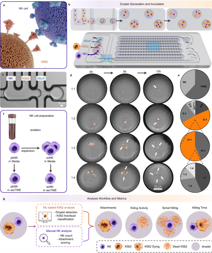
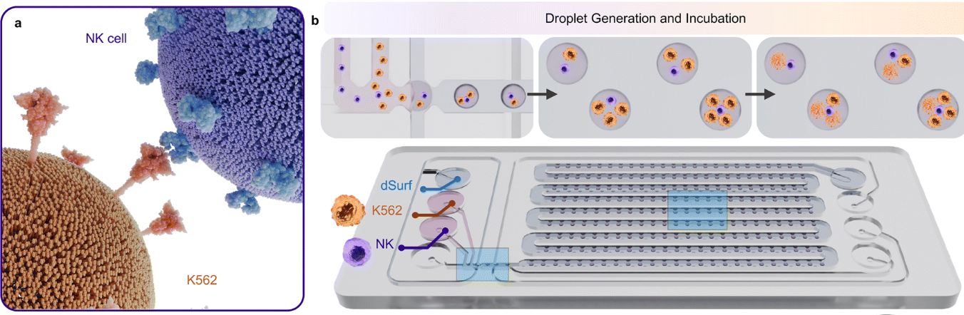
“Single-cell analysis of NK cell cytotoxicity in a droplet microfluidic platform. (a) Illustration depicting NK cell-mediated cytotoxicity against K562 tumor cells. NK cell recognizes and attacks K562 tumor cell through directed cytotoxic mechanisms. (b) Droplet generation and incubation. Top: schematic of the flow-focusing process where NK cells (purple) and K562 target cells (orange) are co-encapsulated with dSurf oil to form droplets. Bottom: microfluidic chip (Fluidic 719) showing droplet array storage for 10-hour time-lapse imaging. (c) Representative brightfield microscopy image of the flow-focusing junction showing the NK, K562, and dSurf inlet streams during droplet generation. Scale bar, 50 μm. (d) Representative time-lapse images showing NK cell interactions with K562 cells over 10 hours at varying effector-to-target (E : T) ratios (1 : 1, 1 : 2, 1 : 3, 1 : 4) using 10× magnification. K562 cells appear as orange fluorescent spots (TRITC overlay). White arrows highlight dead K562 cells. Scale bar, 50 μm. (e) Quantification of droplet contents. Top to bottom: distribution of detected cell types across droplets (NK and K562); distribution of droplet classes including empty droplets (0 : 0), co-encapsulated droplets (E : T), K562-only droplets (0 : T), NK-only droplets (E : 0), and droplets with cell clusters; distribution of E : T droplets by NK cell number (E = 1 for single NK cell, E > 1 for multiple NK cells); and distribution of E = 1 droplets by K562 number from 1 to 4. (f) Schematic illustrating NK cell preparation workflow: isolation from peripheral blood, ex vivo expansion, and conditioning in ascites tumor microenvironment (ascTME) to generate four NK cell states: pbNK, exNK, pbNK-asc, and exNK-asc. (g) Analysis workflow and output metrics derived from droplet time-lapse imaging. K562-related analysis was performed using an ML-based workflow for droplet detection and K562 live/dead classification, whereas NK-related outputs were obtained from morphology-guided NK counting and attachment scoring in the time-lapse images. These complementary analyses were integrated to quantify attachment outcomes, killing activity, serial killing, and killing time.” Reproduced from R. S. Ozcan, F. Vahedi, S. Namakian, A. A. Ashkar and T. F. Didar, Lab Chip, 2026, Advance Article, under a Creative Commons Attribution 3.0 Unported Licence.
The microfluidic device generated and stored droplets on-chip, creating thousands of small reaction chambers for live-cell analysis. NK cells and fluorescent K562 target cells were introduced to the microfluidic chip through separate aqueous inlets, while oil was used as the continuous phase to form droplets. The authors focused their analysis on droplets containing one NK cell and one to four target cells, which made it possible to compare cell-killing behavior across different effector-to-target conditions. After droplet formation, the microfluidic chip was incubated under standard cell culture conditions and imaged over 10 hours.
A key part of the study was combining microfluidics experiments with the use of machine learning to analyze target-cell death. The K562 cancer cells expressed a fluorescent marker, allowing the researchers to follow them during time-lapse imaging. A trained image analysis model detected droplets and classified target cells as alive or dead based on their fluorescence and morphology. This helped convert large microscopy datasets into time-resolved killing profiles for individual NK cells.
The authors compared freshly isolated peripheral blood NK cells, expanded NK cells, and both cell types after exposure to malignant ascites from ovarian cancer patients. This comparison was important because malignant ascites represents an immunosuppressive tumor environment that can reduce NK cell function. The results showed that expanded NK cells were more effective killers than freshly isolated NK cells. They killed a larger fraction of target cells, acted faster, and were more likely to perform serial killing, meaning that a single NK cell could eliminate more than one target cell.
The study also showed how strongly the tumor environment can suppress NK cell activity. Fresh NK cells exposed to malignant ascites showed much weaker target attachment and cytotoxicity. Expanded NK cells were also affected by ascites, but they retained more killing activity than fresh NK cells under the same condition. This suggests that ex vivo expansion may help NK cells maintain some function in suppressive tumor-like environments.
Overall, this work shows how droplet microfluidic devices can provide a detailed view of immune cell function that bulk assays cannot capture. By combining single-cell compartmentalization, live-cell imaging, and machine learning analysis, the authors measured not only whether NK cells killed cancer cells, but also how they killed, how fast they acted, and how their function changed after exposure to an immunosuppressive environment. For microfluidics and cancer immunotherapy research, this study demonstrates the value of droplet-based single-cell assays for evaluating NK cell therapies with much higher functional detail.
Figures are reproduced from R. S. Ozcan, F. Vahedi, S. Namakian, A. A. Ashkar and T. F. Didar, Lab Chip, 2026, Advance Article , DOI: 10.1039/D6LC00301J under a Creative Commons Attribution 3.0 Unported Licence.
Read the original article: Droplet microfluidic profiling of NK cell cytotoxicity with machine learning-enabled target-cell death analysis
For more insights into the world of microfluidics and its burgeoning applications in biomedical research, stay tuned to our blog and explore the limitless possibilities that this technology unfolds. If you need high quality microfluidics chip for your experiments, do not hesitate to contact us.