15 Jun Detecting Antibiotic Resistance with Droplet Microfluidics and Image Texture Analysis
Antibiotic resistance is not always easy to detect. In some bacterial infections, most cells may appear susceptible to an antibiotic, while a very small subpopulation can survive treatment. This phenomenon, called heteroresistance, is clinically important because these rare resistant cells can expand during antibiotic exposure and may contribute to treatment failure. The challenge is that these subpopulations can be extremely rare, often occurring at frequencies around 10⁻⁷ to 10⁻⁴, making them difficult to detect with standard antibiotic susceptibility testing. The current gold-standard population analysis profile test can identify heteroresistance, but it is slow, labor-intensive, and not practical for routine clinical use. In this microfluidic study, the research team addressed this diagnostic gap by developing a droplet microfluidic method for detecting rare antibiotic-resistant subpopulations from bloodstream infection isolates.
The authors combined droplet microfluidics with image texture quantification to create a digital phenotyping approach for heteroresistance detection. Instead of relying only on bulk bacterial growth or single-cell encapsulation, they used a microfluidic chip and loaded bacteria into each droplet and monitored whether any resistant cells could grow under antibiotic pressure. The key idea was that droplets containing growing resistant bacteria develop visible texture changes over time, while droplets without growth remain more uniform. By measuring these texture differences computationally, the microfluidic platform could distinguish growth-positive droplets from no-growth droplets and estimate the frequency of resistant subpopulations. This fluidic approach was tested across both Gram-negative and Gram-positive bloodstream infection isolates, including Acinetobacter baumannii, Klebsiella pneumoniae, Pseudomonas aeruginosa, and Staphylococcus aureus.
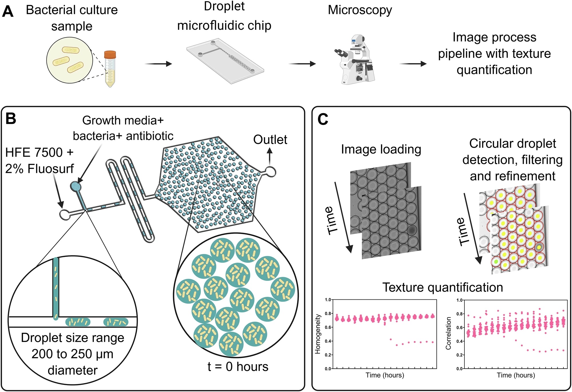
“A The main steps involved in the workflow from overnight bacterial culture to the image process pipeline, involving texture quantification. B Illustration of the microfluidic geometry for droplet generation and storage involving HFE-7500 with 2% Fluosurf as a continuous phase and growth medium with bacteria and the antibiotic of choice as the dispersed phase. C Main steps involving the texture quantification from the time-lapse image sequence. Each dot in a plot of homogeneity and correlation represents the value of texture quantification of every droplet analyzed. Created in BioRender. Agnihotri, S. (2026)” Reproduced from Agnihotri, S.N., Fatsis-Kavalopoulos, N., Vikdahl, E. et al. Droplet microfluidics with image texture quantification for detection of rare antibiotic-resistant subpopulations from bloodstream infections. npj Digit. Med. 9, 410 (2026)., under a Creative Commons Attribution Attribution 4.0 International
The team used a droplet microfluidic device designed for both droplet generation and storage. The continuous oil phase consisted of HFE-7500 with 2% FluoSurf. The dispersed phase contained bacterial culture medium, the antibiotic of interest, and diluted overnight bacterial cultures. The fluids were introduced into the microfluidic chip using syringe pumps, with typical flow rates of 4 µL/min for the oil phase and 1 µL/min for the bacterial suspension. This produced monodisperse droplets with diameters generally around 200 to 250 µm.
After droplet generation, the droplets were kept inside the same microfluidic device and incubated on a microscope stage at 37 °C. The authors acquired time-lapse images to monitor bacterial growth inside droplets. For A. baumannii, K. pneumoniae, and P. aeruginosa, imaging was performed for 24 hours, while S. aureus experiments were followed for 48 hours because the resistant subpopulations appeared more slowly. The workflow shown in the figure summarizes the full process, starting from bacterial culture preparation, moving to the microfluidic device and encapsulation, microscopy, and then image processing.
For image analysis, the authors built a pipeline that segmented droplets and quantified image texture using gray-level co-occurrence matrix features. Among several texture features, homogeneity and correlation were selected as the most useful markers for identifying bacterial growth. Droplets with growing bacteria showed a drop in these texture parameters, while droplets without growth stayed relatively stable. Importantly, the method does not require fluorescent labels or genetic modification. It relies on brightfield microscopy and computational analysis of droplet images.
As a few examples, the proposed microfluidic platform successfully detected heteroresistance in all four bacterial species tested. For A. baumannii exposed to amikacin, the non-heteroresistant strain showed no growth in droplets over 24 hours, while the heteroresistant strain produced many growth-positive droplets. The calculated resistant subpopulation frequencies were around 10⁻⁴, matching the population analysis profile results. For K. pneumoniae, the method again separated non-heteroresistant and heteroresistant strains clearly. The estimated resistant subpopulation frequency was in the 10⁻⁵ to 10⁻⁴ range, depending on the replicate and texture feature used. For P. aeruginosa, the microfluidic platform detected resistant subpopulations, although correlation was less reliable because antibiotic exposure created small residual structures in some droplets, likely from bacterial debris or lysed cells.
Overall, the method detected resistant subpopulations down to approximately one resistant bacterium among one million susceptible cells. The authors reported that the assay could identify growth earlier than the older droplet shrinkage-based readout and faster than the population analysis profile test. They also found that the estimated subpopulation frequencies from texture analysis matched closely with population analysis profile measurements across the tested strains.
This microfluidic study presents a strong example of how a microfluidic device can be paired with image analysis to address a difficult clinical microbiology problem. By confining bacterial populations in droplets and tracking texture changes over time, the platform can detect rare antibiotic-resistant subpopulations from bloodstream infection isolates with high sensitivity. While the method still needs further validation and optimization for clinical use, especially across more antibiotics, strains, and sample types, it offers a practical path toward faster heteroresistance detection.
Figures are reproduced from Agnihotri, S.N., Fatsis-Kavalopoulos, N., Vikdahl, E. et al. Droplet microfluidics with image texture quantification for detection of rare antibiotic-resistant subpopulations from bloodstream infections. npj Digit. Med. 9, 410 (2026). https://doi.org/10.1038/s41746-026-02808-x under a Creative Commons Attribution Attribution 4.0 International
Read the original article: Droplet microfluidics with image texture quantification for detection of rare antibiotic-resistant subpopulations from bloodstream infections
For more insights into the world of microfluidics and its burgeoning applications in biomedical research, stay tuned to our blog and explore the limitless possibilities that this technology unfolds. If you need high quality microfluidics chip for your experiments, do not hesitate to contact us.