


07 Jul Artificial Red Blood Cells for Microfluidic Hemorheology
Blood is difficult to recreate in the lab because its flow behavior is not controlled by plasma alone. Much of its complexity comes from red blood cells, which deform, interact with one another, migrate within vessels, and respond strongly to confinement. This becomes especially important in microcirculation, where vessel dimensions approach the size of individual red blood cells. In these narrow environments, cells no longer behave like passive particles. They stretch, align, form single-file arrangements, and adopt shapes such as parachute-like or bullet-like morphologies.
This creates a challenge for microfluidics. Many blood-mimicking fluids can reproduce certain bulk properties of blood, such as viscosity or shear-thinning behavior, but they often fail to capture single-cell mechanics inside confined microchannels. Some blood analogues use spherical particles, while others focus on matching macroscopic rheology rather than reproducing the size, shape, deformability, and aggregation behavior of real red blood cells. Native blood is also not always ideal for systematic device testing because it varies from donor to donor, changes during storage, and may be difficult to obtain in consistent quantities. For lab-on-a-chip systems, organ-on-chip models, and blood-contacting medical devices, a more standardized and controllable model fluid would be highly useful.
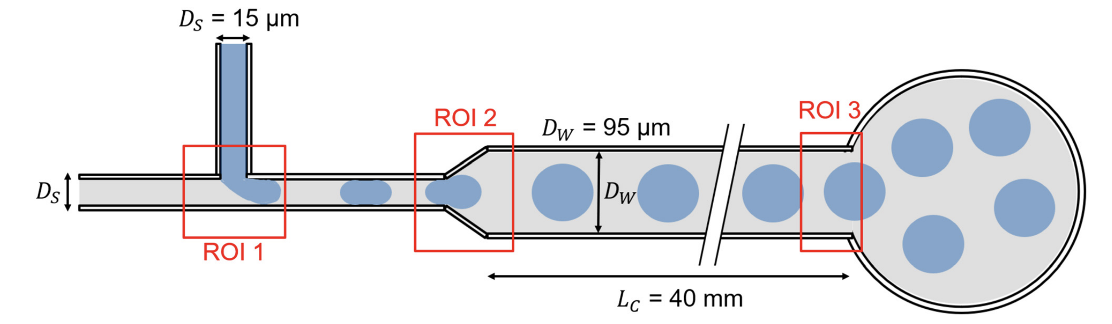
“Schematic of the cross-flow microfluidic device used for particle generation. Hydrogel precursor (blue) is injected as the disperse phase into a mineral oil continuous phase (grey), forming water-in-oil droplets at the cross-junction (ROI 1). As droplets pass through the channel expansion (ROI 2), confinement is reduced, enabling relaxation from plug-like to disc-like morphologies. Subsequent polymerization and removal of the particle core result in the formation of biconcave artificial erythrocytes. The channel dimensions (height 11–15 μm, width expansion from 15 to 95 μm) are designed to match physiological confinement relevant to red blood cells.” Reproduced from Gesine Hentschel, Steffen M. Recktenwald, Katharina Doll-Nikutta, Jan F. Drexler, Maren S. Prediger, Marc Mueller, Marc Wurz, Amy Q. Shen, Birgit Glasmacher; A particulate blood-mimicking fluid with physiological biconcave geometry for microscale hemorheology. Lab Chip 2026; under a Creative Commons Attribution 3.0 Unported License.
The central idea was to build a model system that could capture more than one aspect of blood behavior at the same time. The particles needed to be small enough for capillary-scale confinement, soft enough to deform under flow, shaped more like real erythrocytes than spherical particles, and responsive to different plasma-like environments. By adjusting the suspending medium, the researchers could influence swelling, stiffness, and particle-particle interactions.
The researchers fabricated artificial erythrocytes using a cross-flow microfluidic droplet generator. The microfluidic fabrications were done with PDMS using soft lithography and then plasma-bonded to glass slides. The channel design included a narrow region and a wider expansion region, which helped shape the droplets as they formed and moved through the device. The hydrogel precursor, poly(sodium-acrylate-co-acrylamide), was used as the dispersed phase, while mineral oil containing TEMED served as the continuous phase. This produced water-in-oil droplets that polymerized into hydrogel microparticles.
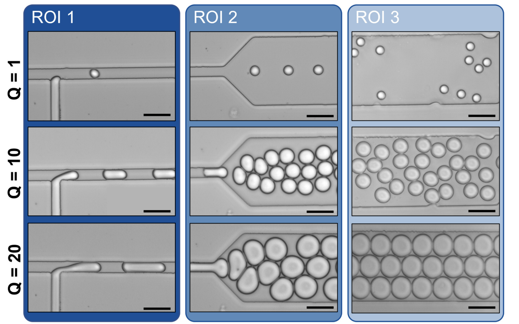
The geometry of the microfluidic channel played a major role in particle formation. As the ratio between the dispersed and continuous phase flow rates changed, the droplets changed in size and shape. At low flow-rate ratios, droplets were closer to spherical and well separated. At higher ratios, they became more elongated and plug-like inside the confined channel. When these droplets entered the wider expansion region, they relaxed toward disc-like shapes. After collection and washing, weakly polymerized material from the particle core was removed, leaving behind a biconcave morphology similar to the shape of red blood cells.
After fabrication, the particles were transferred into different artificial plasma phases. Two of these were glycerol-water mixtures, one containing 10% glycerol and the other 50% glycerol. The third was a dextran 40/CaCl2 solution, chosen because dextran can promote red blood cell-like aggregation behavior. This allowed the authors to compare how the surrounding medium affected particle swelling, stiffness, aggregation, and flow response.
The team then characterized the artificial erythrocytes under static and flowing conditions. They measured particle diameter over time to assess swelling after 24 hours, 48 hours, 72 hours, and 7 days. They studied aggregation by imaging particle suspensions under static conditions and calculating how many particles were present in each aggregate. Mechanical properties were measured using atomic force microscopy in liquid, where individual particles were indented to estimate Young’s modulus and compare deformation behavior across the different suspending media.
To test capillary-scale flow behavior, the researchers pumped dilute particle suspensions through straight microfluidic chips with a 12 μm by 12 μm cross-section. This microchannel size was chosen to approximate strong confinement, similar to the conditions red blood cells experience in small vessels. Particle motion in the microfluidic chips was recorded under microscopy, and image analysis was used to measure length, diameter, projected area, velocity, and deformation index. These measurements allowed the authors to compare how the artificial erythrocytes changed shape as flow velocity increased.
“Representative microscopy images of artificial erythrocyte (ARBC) formation across three regions of interest (ROI 1: cross section, ROI 2: channel width transition, ROI 3: channel outlet) for three exemplary flow rate ratios (Q = 1, 10, 20). The particles are produced with a W/Oemulsion, where the disperse phase is the hydrogel PSAAm and the continuous phase is mineral oil with the added catalyst TEMED. The scale bar corresponds to 30 μm.” Reproduced from Gesine HentschelSteffen M. RecktenwaldKatharina Doll-NikuttaJan F. DrexlerMaren S. PredigerMarc MuellerMarc WurzAmy Q. ShenBirgit Glasmacher; A particulate blood-mimicking fluid with physiological biconcave geometry for microscale hemorheology. Lab Chip 2026; under a Creative Commons Attribution 3.0 Unported License.
The first important result was that the microfluidic fabrication process could produce highly uniform hydrogel particles with biconcave geometry. This is important because many particulate blood analogues rely on spherical particles, which do not reproduce the geometry-dependent deformation behavior of red blood cells. Here, the particles formed reproducibly, and the shape could be influenced by the flow-rate ratio during droplet generation. When the artificial erythrocytes were pumped through confined 12 μm microchannels, they deformed in a velocity-dependent manner. At lower velocities, the particles remained closer to disc-like shapes. At higher velocities, they elongated and adopted bullet-like morphologies in the microfluidic chips. This trend is similar to the behavior of red blood cells under strong confinement, where cell shape depends on flow conditions, confinement, and the properties of the suspending medium.
Overall, the study moves blood-mimicking fluids closer to the single-cell scale that matters most in microcirculation. By combining microfluidic fabrication with hydrogel particle mechanics and plasma-phase control, the authors created a useful microfluidic model system for studying how red blood cell-like particles behave inside narrow channels.
Figures are reproduced from Gesine Hentschel, Steffen M. Recktenwald, Katharina Doll-Nikutta, Jan F. Drexler, Maren S. Prediger, Marc Mueller, Marc Wurz, Amy Q. Shen, Birgit Glasmacher; A particulate blood-mimicking fluid with physiological biconcave geometry for microscale hemorheology. Lab Chip 2026; https://doi.org/10.1039/d6lc00290k under a Creative Commons Attribution 3.0 Unported License.
Read the original article: A particulate blood-mimicking fluid with physiological biconcave geometry for microscale hemorheology
For more insights into the world of microfluidics and its burgeoning applications in biomedical research, stay tuned to our blog and explore the limitless possibilities that this technology unfolds. If you need high quality microfluidics chip for your experiments, do not hesitate to contact us.