


31 Aug Mapping Geometry-Dependent Biocementation in Microfluidic Pore Networks
Microbially induced calcium carbonate precipitation (MICP) uses microorganisms to generate CaCO3 within porous materials, helping bind particles together and increase mechanical integrity. This process has potential applications in soil stabilization, crack sealing, and living building materials. However, the performance of biocemented materials depends not only on how much mineral forms, but also on where it forms and whether deposits remain small and dispersed or grow into larger structures that bridge nearby surfaces. The role of three-dimensional pore geometry in controlling these precipitation patterns is still not fully understood, partly because conventional characterization methods provide limited access to mineral growth at the pore scale.
Filanoski and colleagues developed a microfluidic pore-network platform that allowed them to independently vary several geometric features while keeping the bacterial culture and cementation chemistry consistent. The microfluidic device used arrays of cylindrical pillars as a controlled model of granular porous media, with pillar diameter representing grain size, porosity representing void fraction, and channel height representing the out-of-plane pore dimension. By combining this controlled microfluidic geometry with microscopy and automated image analysis, the researchers could examine how each parameter influenced CaCO3 precipitation.
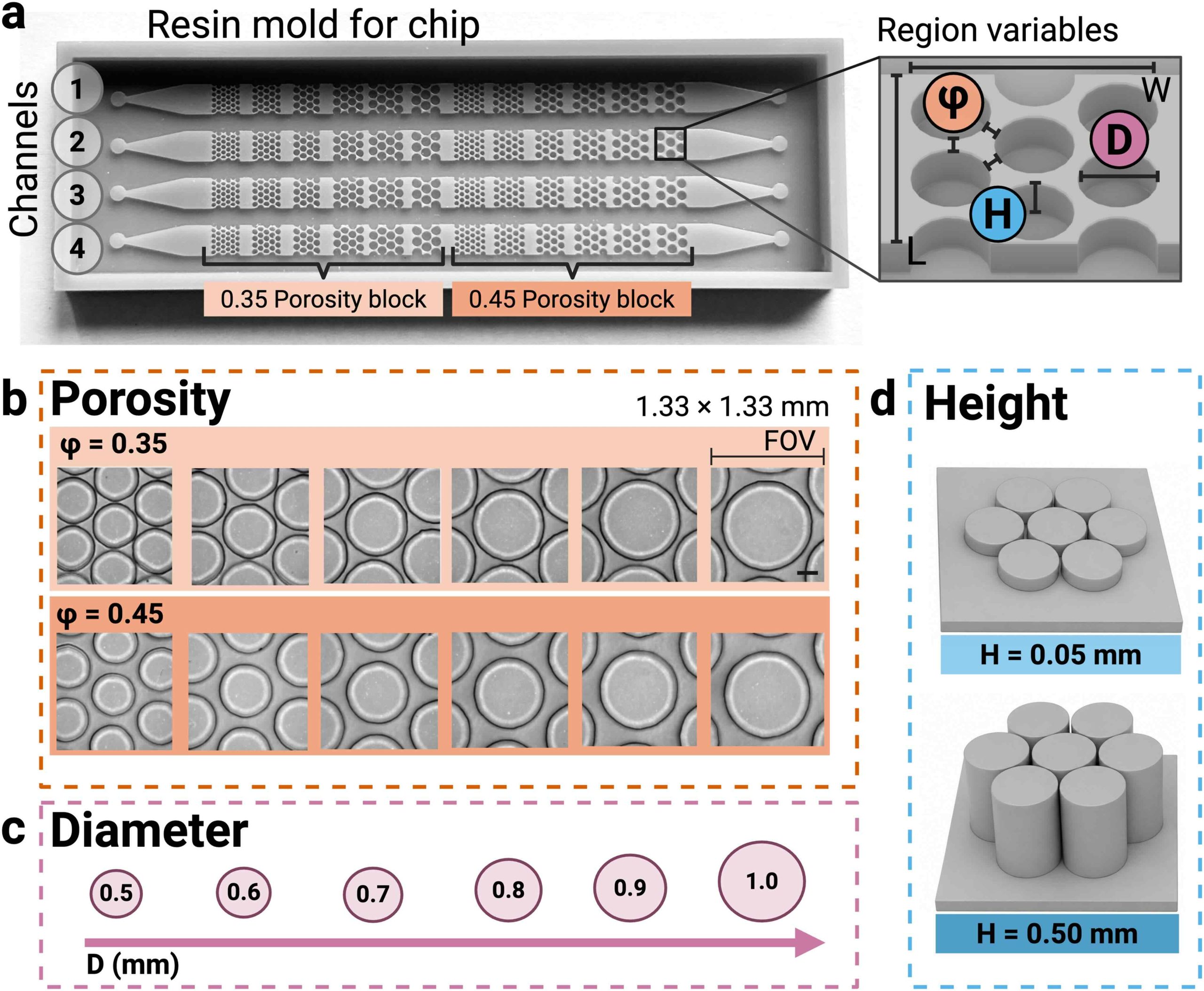
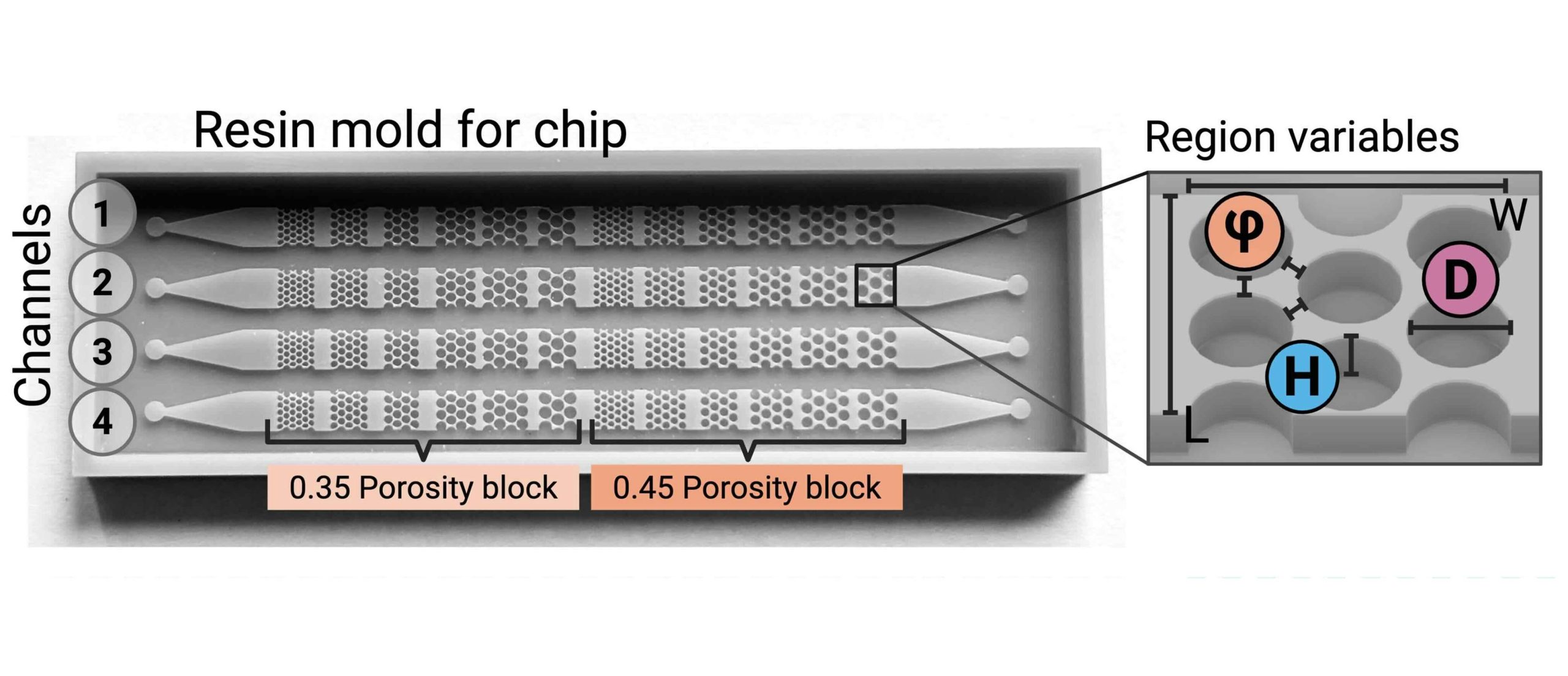
“Overview of pore-network design and device parameters. (a) Resin mold for the chip showing four channels, each divided into 0.35 and 0.45 porosity blocks; inset defines the per-region variables (porosity φ, pillar diameter D, channel height H, field width W, length L). (b) Brightfield fields of view across the six pillar diameters at φ = 0.35 and φ = 0.45; the labeled FOV indicator denotes the 1.33 × 1.33 mm imaged field; scale bar, 250 μm, applies to all panels in (b). (c) Pillar diameters (D = 0.5–1.0 mm). (d) The two channel heights, H = 0.05 mm and H = 0.50 mm.”. Reproduced from Brooke E. FilanoskiMarisa L. BobalKendal R. PhinneyDarke R. HullDavid Erickson; Biocementation shows a three-dimensional geometry dependence in microfluidic pore networks mapped by deep-learning segmentation. Lab Chip 2026; with permission from The Royal Society of Chemistry.
For microfluidics fabrication, the researchers designed the molds in Fusion 360 and produced them using a resin 3D printer with a 20 μm layer thickness. PDMS was cast against the printed molds at a 10:1 base-to-curing-agent ratio, cured, removed from the molds, and plasma-bonded to glass slides. Each microfluidic chip contained four parallel channels divided into regions with porosities of 0.35 and 0.45 and pillar diameters ranging from 0.5 to 1.0 mm. Separate microfluidic chips were fabricated with channel heights of 0.05 and 0.5 mm. The team also produced a single-channel microfluidic device containing 0.10, 0.25, and 0.50 mm height sections to test the effect of channel depth within the same device.
The microfluidic devices were loaded with Sporosarcina pasteurii, a ureolytic bacterium commonly used for MICP, mixed with a cementation solution containing calcium chloride and urea. The microchannels were then maintained under static conditions without imposed flow, allowing precipitation to develop for 16 hours. Brightfield microscopy was used to visualize the pore geometry, while transmitted-light polarization microscopy detected birefringent CaCO3. A U-Net segmentation model trained on manually annotated images automatically identified the pillars, allowing mineral precipitation to be quantified specifically within the open pore space. Microchannel height and porosity had clear effects on mineralization, while pillar diameter showed no measurable effect within the tested range. Lower porosity produced more particles per unit pore area, but those deposits tended to be smaller.
The microfluidic study demonstrates that biocementation in porous networks depends strongly on three-dimensional geometry, particularly microchannel height and porosity. The combination of resin-based mold fabrication, controlled microfluidic devices, polarization imaging, and deep-learning segmentation provides a practical way to study how pore-scale structure influences MICP and may help establish design rules for engineered biocemented materials.
Figures are reproduced from Brooke E. Filanoski, Marisa L. Bobal, Kendal R. Phinney, Darke R. Hull, David Erickson; Biocementation shows a three-dimensional geometry dependence in microfluidic pore networks mapped by deep-learning segmentation. Lab Chip 2026; https://doi.org/10.1039/d6lc00125d with permission from The Royal Society of Chemistry.
Read the original article: Biocementation shows a three-dimensional geometry dependence in microfluidic pore networks mapped by deep-learning segmentation
For more insights into the world of microfluidics and its burgeoning applications in biomedical research, stay tuned to our blog and explore the limitless possibilities that this technology unfolds. If you need high quality microfluidics chip for your experiments, do not hesitate to contact us.