23 Mar Microfabricated structures enable the formation of an in vitro neural network
The study of the human brain, a complex and intricate organ, has always been a frontier in neuroscience. A recent study in this field has been presented in a study published in Lab on a Chip, where researchers have employed a microfluidic chip and construct neuronal circuits using human-induced pluripotent stem cells (iPSCs). Induced pluripotent stem cells (iPSCs) are a type of stem cell that can be generated directly from adult cells. The ability to produce iPSCs revolutionized medical research, offering potential cures for various diseases and an exceptional tool for drug development and personalized medicine.
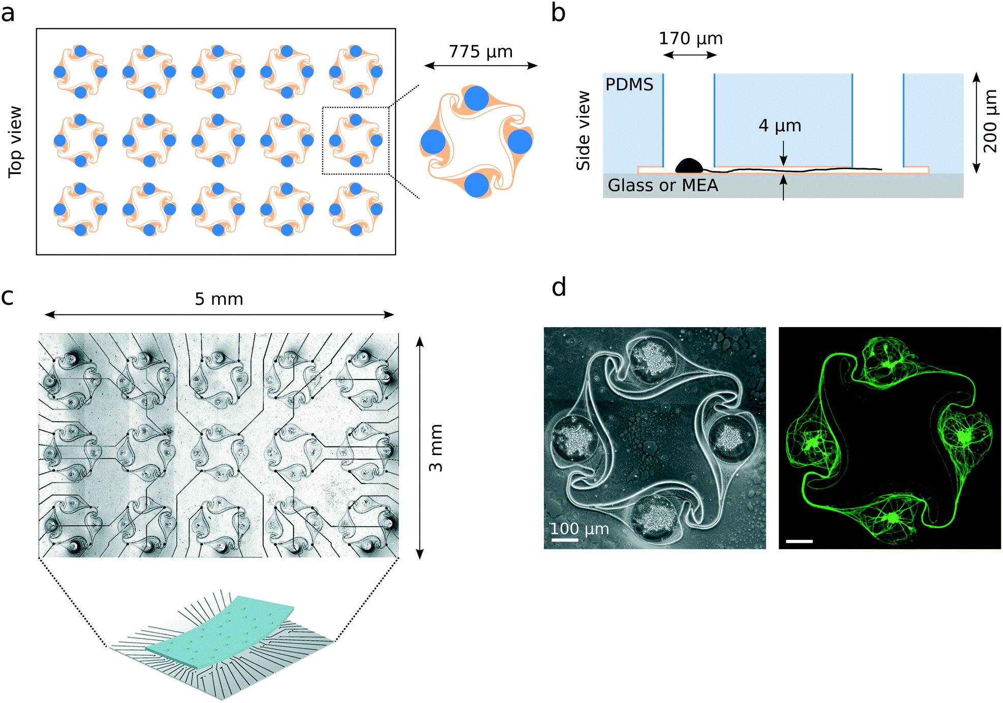
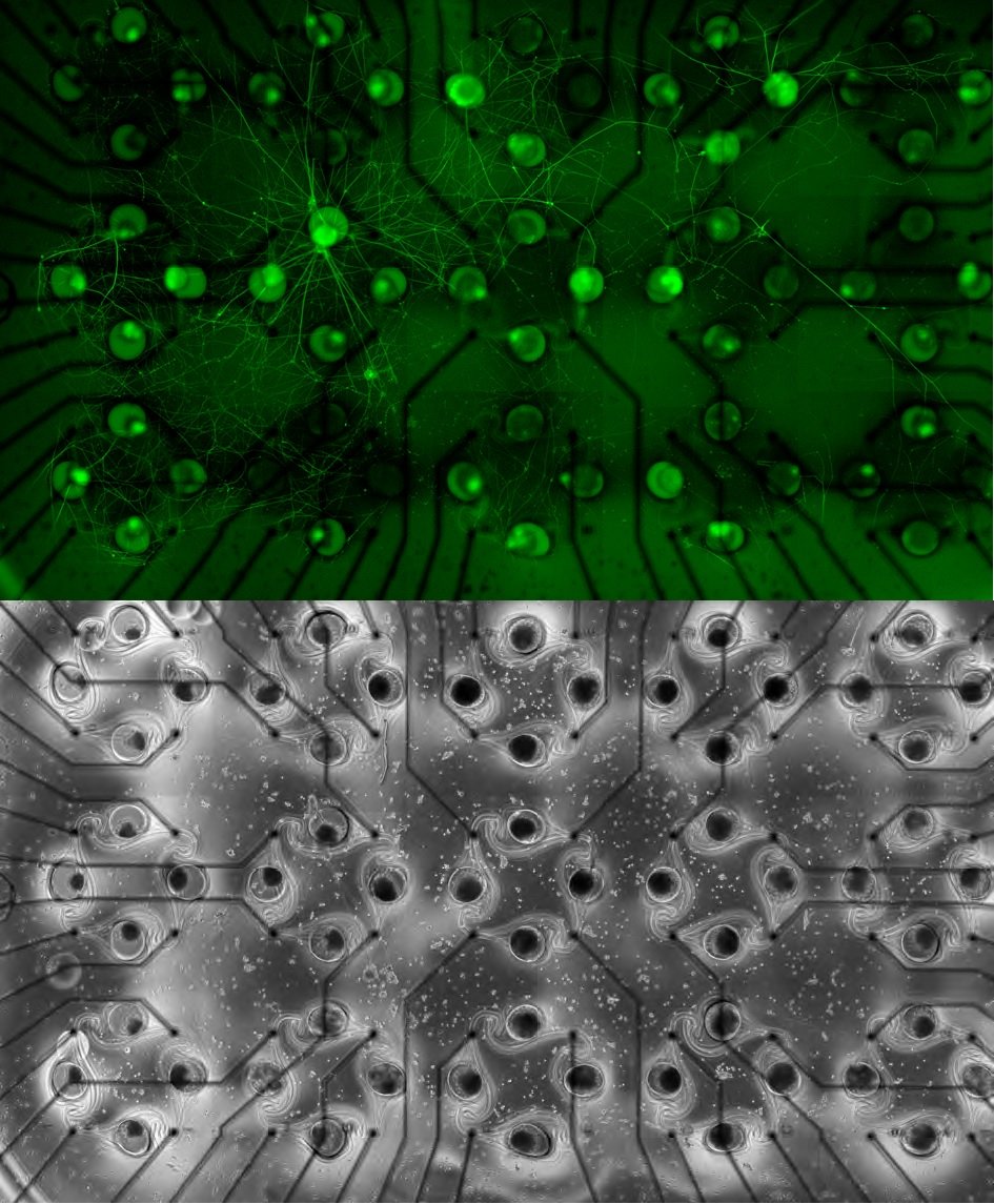
Overview of the PDMS microstructures used to build circuits of iNeurons with controlled axon guidance. (a) Top view of the layout of a typical PDMS microstructure, consisting of 15 circuits, with a zoom-in on one of the circuits. A circuit consists of four nodes (blue) connected by narrow microchannels (orange). The “stomach” shape of the channels allows for axon guidance, resulting in mostly unidirectional, clockwise physical connections between the nodes (see Fig. 5). (b) Schematic side view of two nodes (blue) connected by a microchannel (orange) where an axon is growing [not to scale]. The microchannels are too low for the soma to migrate into, resulting in the physical confinement of the soma in the nodes. (c) Micrograph of a PDMS microstructure with 15 circuits aligned to the 60 electrodes of a MEA. One electrode is positioned under each of the four narrow microchannels of a 4-node circuit, allowing to record from the axon bundle passing on top. (d) Example of a circuit of iNeurons cultured in a PDMS microstructure: phase-contrast (left) and fluorescently labelled iNeurons (right, stained with calcein AM). The soma can be identified as the brighter spots visible in the center of each node. Reproduced under Creative Commons Attribution 3.0 Unported Licence from S. Girardin, B. Clément, S. J. Ihle, S. Weaver, J. B. Petr, J. C. Mateus, J. Duru, M. Krubner, C. Forró, T. Ruff, I. Fruh, M. Müller and J. Vörös, Lab Chip, 2022, Advance Article.
The study’s significant achievement was the demonstration of controlled neuronal interactions in a lab setting using microfluidic technology. Electrophysiology recordings from these circuits provided novel insights into neuronal communication, crucial for understanding brain information processing and memory storage. This approach opens new pathways in neuroscience research, offering a more profound understanding of brain functionality and potential applications in studying various neurological disorders. By replicating brain circuits using iPSC-derived neurons employing microfluidics, this study marks a pivotal advancement in neuroscience, bridging the gap between biological brain functions and their synthetic counterparts in the lab.
Figures and the abstract are reproduced from S. Girardin, B. Clément, S. J. Ihle, S. Weaver, J. B. Petr, J. C. Mateus, J. Duru, M. Krubner, C. Forró, T. Ruff, I. Fruh, M. Müller and J. Vörös, Lab Chip, 2022, Advance Article , DOI: 10.1039/D1LC01110C under Creative Commons Attribution 3.0 Unported Licence.
Read the original article: Topologically controlled circuits of human iPSC-derived neurons for electrophysiology recordings