


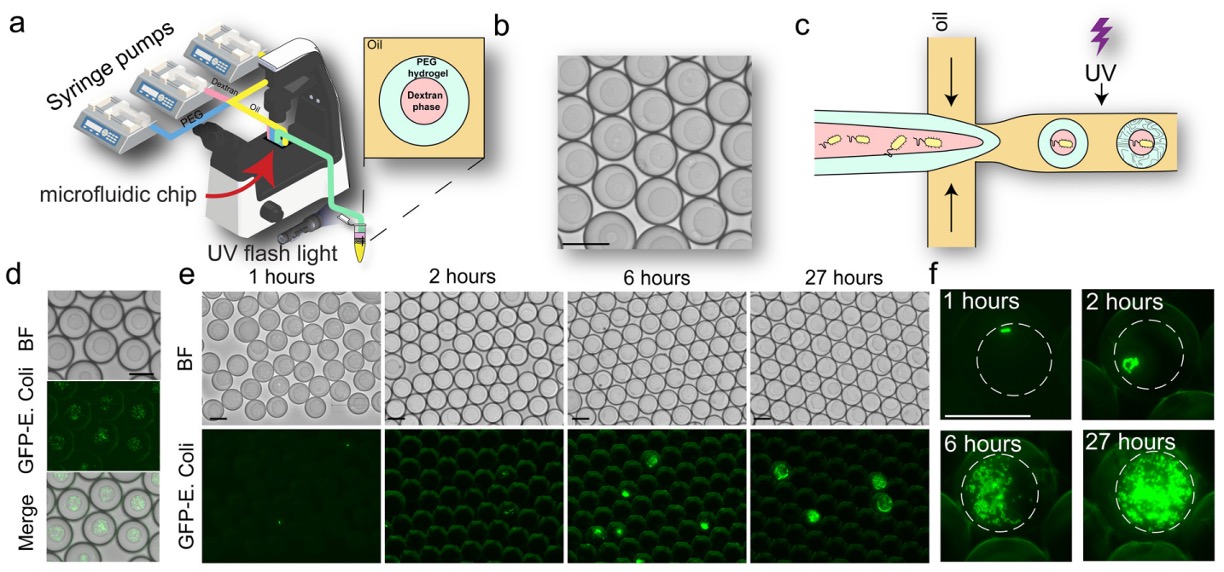
03 Aug Multiphasic Droplet Microfluidics for Controlled Bacteria and Mammalian Cell Co-Culture
Studying interactions between bacteria and mammalian cells is important for understanding the microbiome, infection, inflammation, and other biological processes. However, recreating these interactions in vitro is challenging because the two cell types have very different culture requirements. Mammalian cells often need an adhesive matrix, while bacteria typically grow in liquid media. Existing co-culture systems may also provide limited control over whether the cells interact through direct physical contact or only through secreted molecules.
In this study, researchers developed a multiphasic droplet microfluidics platform that combines liquid and hydrogel compartments within individual droplets. The liquid compartment provides an environment for bacterial growth, while the hydrogel compartment supports mammalian cell adhesion. By changing the internal droplet architecture, the platform can reproduce both direct co-culture, where bacteria and mammalian cells can make physical contact, and indirect co-culture, where they remain physically separated within the same general microfluidic workflow.
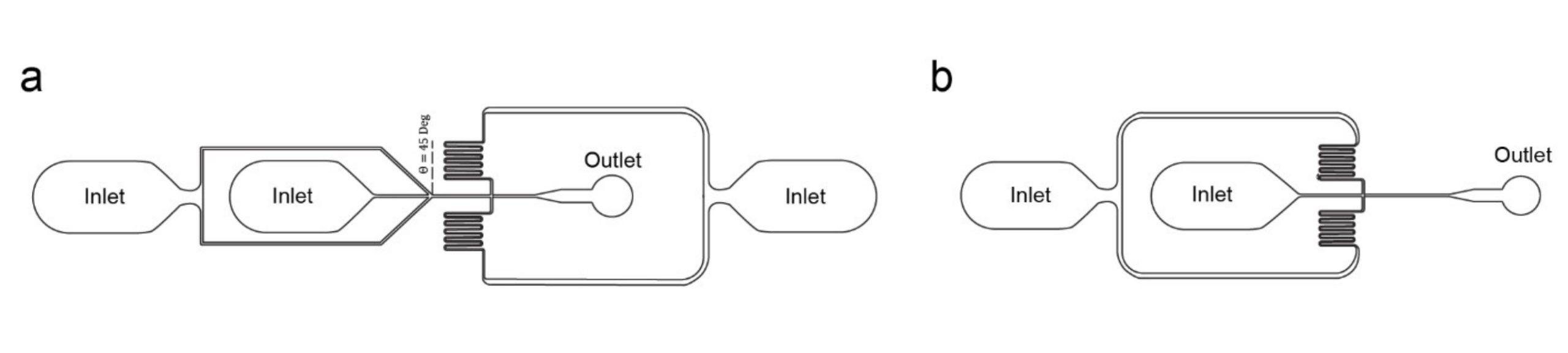
“Design of microfluidic chips for phase separation-based co-encapsulation (a) and sequential encapsulation (b)”. Reproduced from Ibraheem Alshareedah, Anand Kumar; Multiphasic droplet microfluidics platform for controlled bacteria and mammalian cell co-culture. Lab Chip 2026; 26 (8): 2473–2485. with permission from The Royal Society of Chemistry.
Using custom microfluidic chips manufactured by our team at uFluidix, the researchers generated core-shell droplets from polyethylene glycol, or PEG, dextran, and a fluorinated carrier oil. Separate PEG and dextran streams were introduced into a co-flow focusing geometry using syringe pumps. Because the two polymers separate into distinct aqueous phases, the process produced droplets with a dextran-rich liquid core and a PEG-rich outer compartment. Ultraviolet exposure was then used to crosslink the PEG phase into a stable hydrogel.
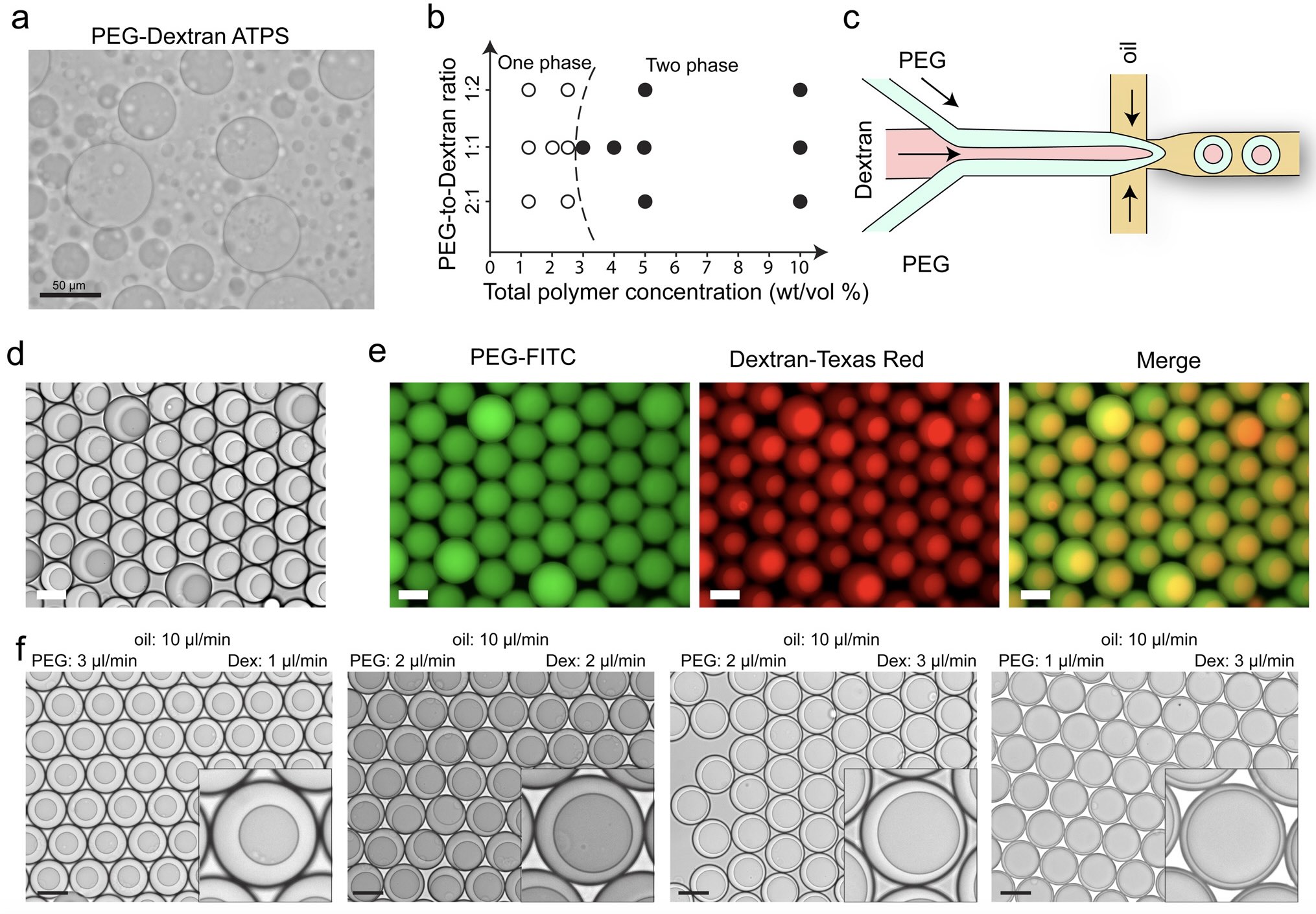
“A microfluidic platform to generate monodisperse multiphasic droplets. (a) Image of the phase separation of a mixture of PEG and dextran. (b) Phase diagram of PEG–DEX mixtures. (c) Diagram of the microfluidic device to create composite core–shell droplets. (d) Image of the microfluidic-generated multiphasic droplet suspension. (e) Fluorescence image showing the partition of both PEG and DEX molecules in MMDs. (f) Bright field images showing the change in multiphasic droplet structure upon changing the relative flow rates of the PEG and dextran phases in their respective channels. Inset shows zoomed-in images. Scale bars represent 50 μm”. Reproduced from Ibraheem Alshareedah, Anand Kumar; Multiphasic droplet microfluidics platform for controlled bacteria and mammalian cell co-culture. Lab Chip 2026; 26 (8): 2473–2485. with permission from The Royal Society of Chemistry.
For direct co-culture, GFP-labelled Escherichia coli Nissle 1917 bacteria were suspended in the dextran-rich phase, while A549 human lung cells were introduced in the PEG hydrogel precursor. The bacteria remained concentrated in the liquid core, and the mammalian cells generally positioned themselves at the boundary between the liquid and hydrogel compartments. This arrangement allowed the mammalian cells to remain associated with an adhesive matrix while also being exposed to the nearby bacterial population.The bacteria continued growing for up to 27 hours without bursting the droplets or entering the PEG-rich hydrogel compartment.
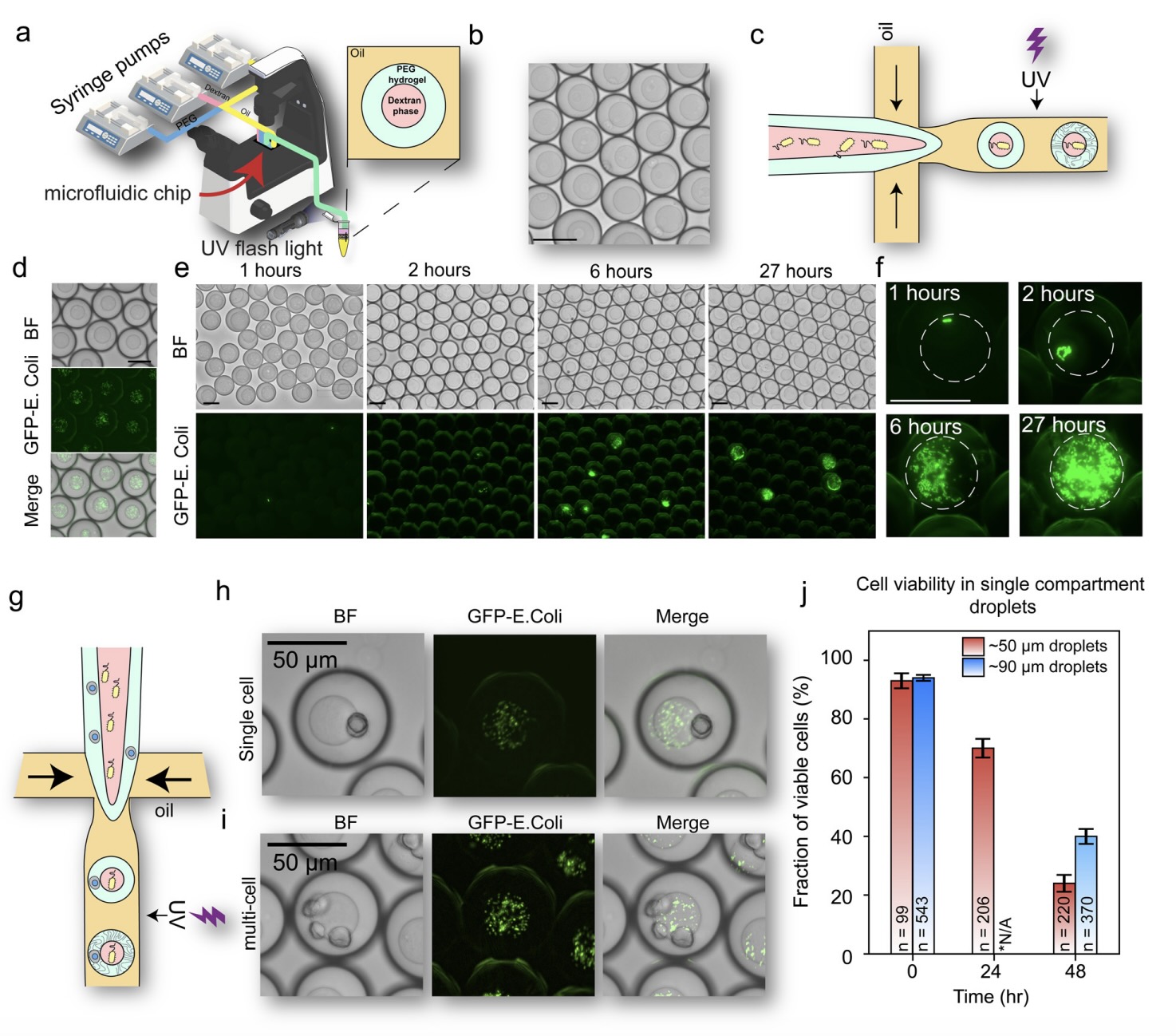
“Co-capture of mammalian cells and bacteria in multi-phasic droplets with a hydrogel shell. (a) Scheme of the experimental setup to create hydrogel MMDs. (b) Brightfield image of MMDs after UV-induced crosslinking. (c) Scheme of bacterial encapsulation in MMDs’ core. (d) Brightfield and fluorescence images of GFP-labeled E. coli encapsulated in the core of MMDs. (e) Brightfield and fluorescence images of the growth of GFP-labeled E. coli in the core of MMDs for 27 hours. (f) Zoomed-in fluorescence images of bacterial colonies at 4 time points within MMDs. The white circle highlights the core of the MMD. (g) Scheme describing the co-encapsulation of bacteria and mammalian cells in MMDs’ core and shell, respectively. (h) Brightfield and fluorescence images of a single cell encapsulated with bacteria in an MMD. (i) Same as (h) but with multi-cell encapsulation. (j) Cell viability plot for cells encapsulated in single-compartment hydrogel droplets of different diameters suspended in oil”. Reproduced from Ibraheem Alshareedah, Anand Kumar; Multiphasic droplet microfluidics platform for controlled bacteria and mammalian cell co-culture. Lab Chip 2026; 26 (8): 2473–2485. with permission from The Royal Society of Chemistry.
The researchers also created an indirect co-culture format using sequential encapsulation. Mammalian cells were first enclosed inside approximately 30 µm PEG hydrogel droplets produced using a chip with a 20 µm nozzle. After ultraviolet curing and transfer into an aqueous solution, these hydrogel particles were re-encapsulated with bacteria using a second chip with a 40 µm nozzle. The resulting droplets were approximately 60 to 70 µm in diameter, with mammalian cells protected inside the hydrogel core and bacteria located in the surrounding liquid compartment. These droplets maintained both cell types for at least 24 hours while preventing direct physical contact. This physical separation prevented direct contact while still allowing metabolites and other small molecules to move between the two regions.
The study also highlighted the balance between droplet size, cell viability, and compatibility with fluorescence-based sorting. Mammalian cell viability in approximately 50 µm droplets decreased from about 93% initially to 70% after 24 hours, while larger droplets provided better survival over longer periods. At the same time, keeping the droplets below roughly 70 to 80 µm makes them more compatible with fluorescence-based sorting systems. Overall, this platform provides a configurable method for examining how physical contact, metabolites, and signalling molecules influence bacteria-mammalian cell interactions at high throughput.
Figures are reproduced from Ibraheem Alshareedah, Anand Kumar; Multiphasic droplet microfluidics platform for controlled bacteria and mammalian cell co-culture. Lab Chip 2026; 26 (8): 2473–2485. https://doi.org/10.1039/d6lc00016a with permission from The Royal Society of Chemistry.
Read the original article: Multiphasic droplet microfluidics platform for controlled bacteria and mammalian cell co-culture
For more insights into the world of microfluidics and its burgeoning applications in biomedical research, stay tuned to our blog and explore the limitless possibilities that this technology unfolds. If you need high quality microfluidics chip for your experiments, do not hesitate to contact us.