


18 Oct ALS-on-a-Chip: Restoring Neuromuscular Signaling by Targeting SLITRK2
Amyotrophic lateral sclerosis (ALS) is a progressive neurological disease in which motor neurons gradually lose their ability to communicate with muscles. Evidence suggests that damage can begin in the distant ends of axons and at neuromuscular junctions (NMJs), where nerves connect to muscle, before motor neurons die. However, the molecular changes responsible for these early defects remain poorly understood. Mutations in FUS, a protein involved in RNA regulation, are associated with ALS, but conventional cell cultures make it difficult to study RNA changes within axons separately from those in neuronal cell bodies.
To address this challenge, a research team combined microfluidics with human induced pluripotent stem cell (iPSC) technology. Their ALS microfluidic model brought together motor neurons carrying ALS-associated FUS mutations and human stem cell-derived skeletal muscle cells to recreate the neuromuscular junction in a controlled environment. In parallel, the team cultured motor neurons in a compartmentalized microfluidic device, making it possible to collect RNA from axons separately and investigate molecular changes associated with the disease.
For microfluidics fabrication, the researchers cast polydimethylsiloxane (PDMS) chambers using a 10:1 mixture of silicone base and curing agent, cured them overnight at 60 °C, and bonded them to glass coverslips after plasma treatment. Each microfluidic chip contained two cell culture compartments connected by narrow microgrooves that guided growing motor neuron axons toward the muscle compartment while keeping neuronal cell bodies separate. The team seeded iPSC-derived muscle progenitors into one microfluidic compartment and induced them to form myotubes, then introduced motor neurons into the other compartment. Over subsequent days, axons extended through the connecting passages and established nerve-to-muscle contacts.
The researchers first collected RNA independently from the axonal and cell-body-containing compartments and analyzed it using CEL-seq2 RNA sequencing. They compared motor neurons derived from patients with FUS-related ALS against healthy and genetically matched controls, examining both gene expression and differences in the untranslated regions of RNA transcripts. After identifying SLITRK2 as a candidate gene, they used lentiviral short hairpin RNA to reduce its expression and tested the effects on axonal growth and neuromuscular signaling. To measure communication between neurons and muscle cells, they stimulated motor neurons with potassium chloride and tracked calcium signals in both cell types using live fluorescence imaging.
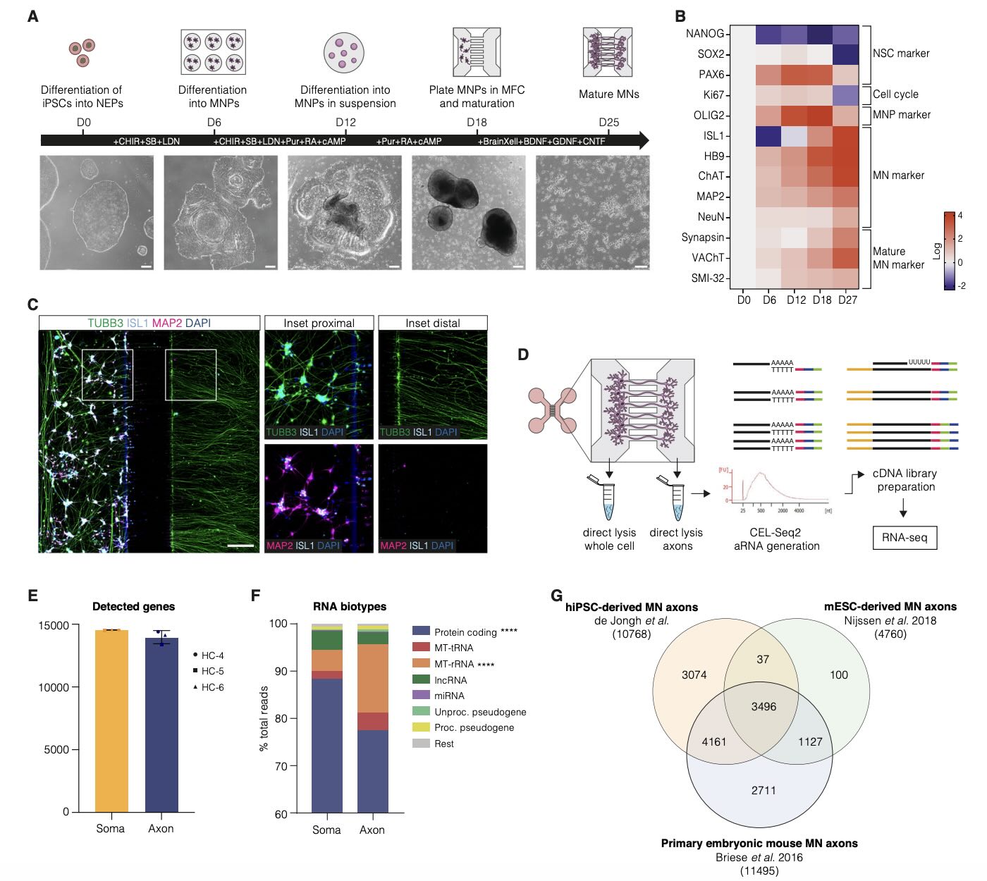
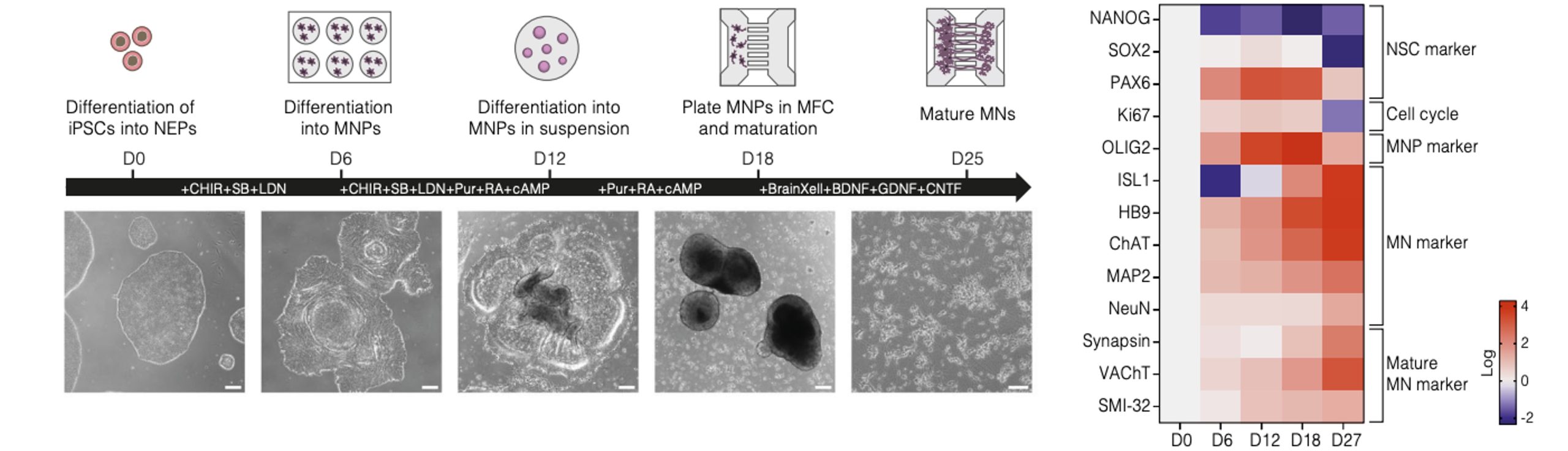
“Generation and characterization of iPSC-derived motor neurons for axonal transcriptomic analysis. A Schematic showing the differentiation protocol of iPSCs into spinal MN, including subsequent culture within microfluidic devices. Representative brightfield pictures illustrate morphology at the indicated time points. Scale bar D0-D18: 200 μm, D25: 100 μm. B mRNA expression levels of several developmental and mature (neuronal) markers at different time points during differentiation (n = 1). Expression levels are normalized to the housekeeping genes GAPDH and β-actin. C Expression of MN somatodendritic markers ISL1 and MAP2, and neurite marker TUBB3 in iPSC-derived MN cultured in microfluidic devices at D27. Scale bar: 200 μm. Insets: magnification of somatodendritic (proximal) and axonal (distal) compartments showing ISL1, MAP2 and TUBB3 expression. D Schematic illustrating the workflow for RNA isolation and sequencing from somatodendritic (whole cell) and axonal compartments of iPSC-derived MN. RNA was directly lysed from each compartment, followed by aRNA generation and cDNA preparation according to the CEL-seq2 protocol prior to RNA-seq. E Number of detected genes in somatodendritic and axonal samples. Data is presented as mean ± SD (n = 3 biological replicates, t = 1 technical replicate). Paired two-tailed Ttest (p = 0.1903). F RNA biotype classification in somatodendritic and axonal samples. Data is presented as mean (n = 3 biological replicates, t = 1 technical replicate). Two-way ANOVA. **** p < 0.0001. G Venn diagram comparing the overlap between genes identified in our human iPSC-derived MN axons compared to those found in previously published datasets from mouse embryonic stem cell-derived MN axons and primary embryonic mouse MN axons”. Reproduced from de Jongh, R., Wiersema, A.F., van Dijk, C.H. et al. SLITRK2 knockdown rescues axonal and functional neuromuscular junction deficits in human iPSC-derived mutant FUS-ALS motor neurons. Nat Commun (2026)., under Creative Commons Attribution 4.0 International License.
The analysis revealed that SLITRK2 was consistently elevated in axons from multiple FUS-ALS models. Mutant motor neurons also displayed abnormally increased axonal outgrowth, which was normalized when SLITRK2 expression was reduced. More importantly, muscle cells connected to FUS-ALS motor neurons showed weaker responses to neuronal stimulation. Lowering SLITRK2 restored these muscle calcium responses, while increasing SLITRK2 in healthy neurons impaired neuromuscular signaling. Interestingly, SLITRK2 knockdown did not restore the reduced excitability of the motor neurons or increase the number of innervated neuromuscular junctions, suggesting that it improved signaling through existing connections rather than creating new ones.
This study demonstrates how a compartmentalized microfluidic human neuromuscular model can connect changes in axonal gene regulation with functional defects in ALS. By combining stem cell-derived motor neurons and muscle cells with targeted gene manipulation and live imaging, the microfluidic platform offers a useful way to investigate early disease mechanisms and evaluate potential interventions.
Figures are reproduced from de Jongh, R., Wiersema, A.F., van Dijk, C.H. et al. SLITRK2 knockdown rescues axonal and functional neuromuscular junction deficits in human iPSC-derived mutant FUS-ALS motor neurons. Nat Commun (2026). https://doi.org/10.1038/s41467-026-78419-7 under Creative Commons Attribution 4.0 International License.
Read the original article: SLITRK2 knockdown rescues axonal and functional neuromuscular junction deficits in human iPSC-derived mutant FUS-ALS motor neurons
For more insights into the world of microfluidics and its burgeoning applications in biomedical research, stay tuned to our blog and explore the limitless possibilities that this technology unfolds. If you need high quality microfluidics chip for your experiments, do not hesitate to contact us.