


06 Nov Sorting Bacterial Cocultures Using Microfluidic Droplets and Photogelation
Bacteria rarely live in isolation. Within the human microbiome, different species compete, cooperate, and influence one another, making it difficult to identify which interactions shape a microbial community. Droplet microfluidics offers a way to study these interactions by confining small groups of bacteria in thousands of separate compartments. However, selecting and recovering droplets with interesting outcomes often requires specialized fluorescence-activated droplet sorting equipment. Encapsulating bacteria in solid hydrogels can make conventional flow sorting possible, but the gel matrix may also restrict bacterial growth. Researchers therefore need a way to combine liquid-phase bacterial culture with straightforward sorting and recovery.
In a study published in Lab on a Chip, researchers at Los Alamos National Laboratory developed a method called post-assay photogelation. Their microfluidic approach keeps bacteria in liquid water-in-oil droplets during the culture period, then converts the droplets into solid hydrogel particles only after the bacterial interactions have taken place. This is achieved by including light-reactive polymer precursors in the microfluidic droplets and exposing them to ultraviolet light after incubation. Once solidified, the particles can be transferred from oil into an aqueous solution, sorted with a commercial flow cytometer, and broken down enzymatically to retrieve the enclosed bacteria.
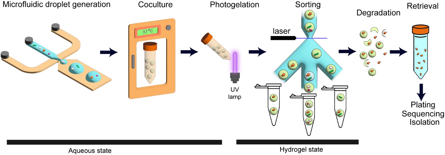
“A droplet microfluidic platform for encapsulation, culture, sorting, and retrieval of bacterial cells.”. Reproduced from Ibraheem AlshareedahKatelyn M. GreenSang-Min ShinRamesh K. JhaAnand Kumar; Post-assay photogelation enables flow-cytometric sorting and recovery of bacterial cocultures from microfluidic droplets. Lab Chip 2026; with permission from The Royal Society of Chemistry.
The researchers used custom-made microfluidic chips manufactured by uFluidix to generate the droplets. One microfluidic device incorporated a cross-junction with a 20 µm nozzle, while a flow-focusing design was also used in the sorting experiments. A bacterial suspension containing hydrogel precursors flowed through the microfluidic devices as the dispersed phase, and HFE7500 oil containing a fluorosurfactant served as the continuous phase. For the cross-junction setup, the team typically used an oil flow rate of 20 µL/min and an aqueous flow rate of 2 to 5 µL/min, producing droplets around 30-40 µm in diameter. The microfluidic droplets were incubated at 37 °C for approximately 18 hours before light-triggered gelation.
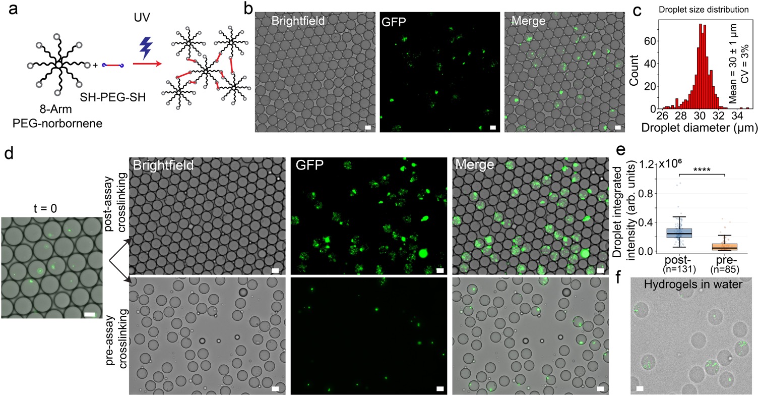
The choice of hydrogel material proved important. Initial experiments with polyethylene glycol (PEG)-based precursors showed that bacteria grown in liquid droplets before gelation had stronger and more evenly distributed fluorescence than bacteria cultured inside gels formed at the start. The team then tested an enzyme-cleavable peptide crosslinker, but this formulation inhibited the growth of the tested Escherichia coli Nissle 1917 strain. Switching to methacrylated dextran produced a light-responsive formulation that supported bacterial growth and could later be degraded with dextranase. After incubation, approximately 60 seconds of ultraviolet exposure stabilized the droplets as particles suitable for transfer into an aqueous carrier and downstream sorting.
“PEG hydrogel formulation with post-assay photogelation to culture bacterial cells. (a) Diagram showing the process of UV crosslinking of 8-arm PEG-norbornene and PEG dithiol as a crosslinker. (b) Brightfield and fluorescence images of GFP-expressing EcN cells encapsulated in microfluidic droplets. (c) Histogram of droplet diameters as measured by microscopy. The mean diameter is 30 ± 1 μm. The error represents one standard deviation. (d) Brightfield and fluorescence images of droplets encapsulating EcN cells along with gel precursor solutions at t = 0 and at t = 18 hours in two conditions: pre-assay crosslinking (crosslinked right after encapsulation), and post-assay photogelation (crosslinked after 18 hours of growth). (e) Integrated GFP intensity per droplet for post-assay crosslinked and pre-assay crosslinked samples, designated as post- and pre-, respectively. The “****” symbol denotes a p-value < 0.0001 from a two-sided Welch’s t-test. The box represents the interquartile range (IQR) while the whiskers represent 1.5× IQR. The black line represents the median and the points are individual values. Each point in the plot represents a droplet and is treated as a technical replicate. (f) EcN-laden hydrogels after transferring them to an aqueous carrier phase for sorting. All scale bars represent 20 μm.”. Reproduced from Ibraheem AlshareedahKatelyn M. GreenSang-Min ShinRamesh K. JhaAnand Kumar; Post-assay photogelation enables flow-cytometric sorting and recovery of bacterial cocultures from microfluidic droplets. Lab Chip 2026; with permission from The Royal Society of Chemistry.
To assess sorting performance, the team first generated red and green fluorescent bead-containing hydrogel particles and separated them using a commercial WOLF G2 cell sorter. The sorted populations reached approximately 99.9% purity for green particles and 99.7% for red particles. In a second experiment, sorting increased the proportion of hydrogel particles containing fluorescent E. coli Nissle from 59% in the input mixture to 98% in the collected sample. A separate recovery test showed that about 69 ± 9% of culturable bacteria could be recovered across the encapsulation, gelation, transfer, and enzymatic digestion workflow. These results demonstrate that the droplets can be converted into sortable particles without permanently trapping the bacteria.
Finally, the researchers used the microfluidic platform to investigate interactions between fluorescent E. coli Nissle and a cultured bacterial community obtained from human nasal samples. After coculture, E. coli Nissle was the predominant detectable population in droplets containing the fluorescent strain, suggesting that it may have inhibited members of the nasal community under the conditions tested. Overall, post-assay photogelation offers a practical way to link aqueous bacterial coculture with conventional particle sorting and subsequent microbial recovery. The study also highlights how custom microfluidic devices can support the development of more accessible bacterial screening workflows.
Figures are reproduced from Ibraheem AlshareedahKatelyn M. GreenSang-Min ShinRamesh K. JhaAnand Kumar; Post-assay photogelation enables flow-cytometric sorting and recovery of bacterial cocultures from microfluidic droplets. Lab Chip 2026; https://doi.org/10.1039/d6lc00749j with permission from The Royal Society of Chemistry.
Read the original article: Post-assay photogelation enables flow-cytometric sorting and recovery of bacterial cocultures from microfluidic droplets
For more insights into the world of microfluidics and its burgeoning applications in biomedical research, stay tuned to our blog and explore the limitless possibilities that this technology unfolds. If you need high quality microfluidics chip for your experiments, do not hesitate to contact us.