


20 Nov Tumor-on-a-Chip Reveals How Hypoxia and Lipid Accumulation Suppress NK Cells
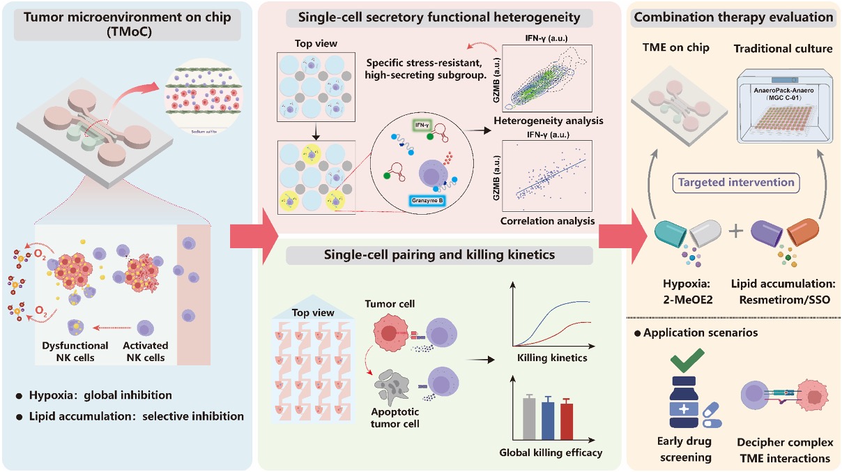
Natural killer (NK) cells help the immune system recognize and destroy cancer cells, but their effectiveness often declines inside solid tumors. Two common features of the tumor microenvironment are hypoxia, or low oxygen availability, and excess lipid accumulation. Both can impair immune responses, yet their individual effects are difficult to distinguish using conventional cell cultures. Two-dimensional models cannot fully reproduce the spatial organization and metabolic stresses of a tumor, while population-level assays hide important differences between individual immune cells. In a recent study published in Lab on a Chip, Aoling Wang and colleagues used microfluidics to investigate how these two conditions affect NK cell activity and whether targeting them together can improve tumor cell killing.
The researchers developed a four-channel tumor-on-a-chip microfluidic chip with a central three-dimensional tumor culture chamber, adjacent fluid channels, and an independent channel for oxygen removal. For microfabrication, they created SU-8 molds using photolithography, cast PDMS at a 10:1 base-to-curing-agent ratio, cured it at 65 °C for two hours, and plasma-bonded the patterned polymer to glass. The central chamber contained Hep3B liver cancer cells in a Matrigel-based three-dimensional matrix, while endothelial EA.hy926 cells formed a vessel-like lining in an adjacent channel. NK-92MI cells were incorporated into tumor co-cultures to assess cytotoxicity or introduced through the endothelial-lined channel in separate experiments to track their infiltration into the tumor region.
“Tumor-on-a-chip with single-cell microfluidics reveals distinct NK cell immunosuppression by hypoxia versus lipid accumulation and enables targeted therapeutic intervention.” Reproduced from Aoling Wang, Yunting Wang, Xiahe Han, Qiurui Chen, Wu Liu; Tumor-on-a-chip with single-cell microfluidics reveals distinct NK cell immunosuppression by hypoxia versus lipid accumulation. Lab Chip 2026; 26 (21): 5791–5804; with permission from The Royal Society of Chemistry.
To create hypoxia, the team introduced a 6-12% sodium sulfite solution into the oxygen-removal channel. A 100 µm-thick, gas-permeable PDMS membrane separated this solution from the culture channels, allowing oxygen depletion without directly mixing the sulfite solution with the cells. To model lipid accumulation, Hep3B cells were pretreated with oleic acid for 24 hours before being loaded into the chip. The researchers then observed NK cell migration, tumor cell viability, and gene expression under normal, hypoxic, lipid-rich, and combined-stress conditions. They also used a separate droplet microfluidics platform to isolate individual NK cells in droplets containing fluorescent sensors for interferon-gamma (IFN-γ) and granzyme B secretion. Another microfluidic device paired single NK cells with single tumor cells, allowing the team to monitor the timing of cancer cell death over 12 hours.
Both hypoxia and lipid accumulation reduced NK cell infiltration and their ability to kill tumor cells, with the strongest impairment observed when the two stresses were combined. However, the underlying responses were different. Hypoxia broadly reduced the expression of molecules involved in NK cell function, including IFN-γ and granzyme B. Lipid accumulation had a more selective effect, reducing granzyme B and IL-10 expression while leaving IFN-γ gene expression relatively unchanged. The single-cell droplet measurements revealed an important distinction: despite the limited change in IFN-γ gene expression under lipid-rich conditions, IFN-γ protein secretion still decreased across the population. A subset of NK cells nevertheless retained relatively high IFN-γ secretion under lipid stress, whereas hypoxia suppressed this response more uniformly.
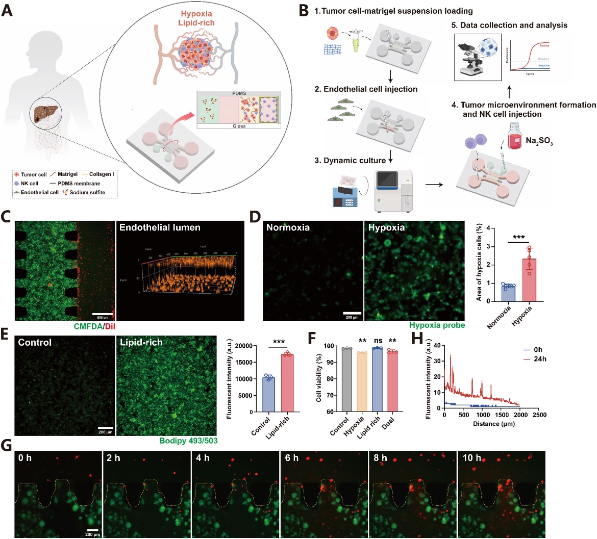
“Establishment and validation of a tumor-on-a-chip model with hypoxic and/or lipid-rich microenvironment. (A) Schematic illustration of the microfluidic device architecture. (B) Scheme of the experimental workflow. (C) Confocal microscopy image showing the spatial distribution of tumor cells (green) and the formation of endothelial lumens (red). (D) Visualization and corresponding quantification of the hypoxia level within the tumor chamber induced by 12% Na2SO3 using the Green Hypoxia Image-iT™ probe. Data are presented as mean ± SD (n = 5). (E) Visualization and corresponding quantification of intracellular lipid droplets induced by 800 μM OA using BODIPY™ 493/503 staining. Data are presented as mean ± SD (n = 3). (F) Viability of tumor cells under the specified culture conditions: control, hypoxia, lipid-rich, and dual stress (hypoxia + lipid-rich). Data are presented as mean ± SD (n = 3). (G) Time-lapse migration and infiltration of NK cells into the tumor chamber within 10 h. Fluorescent micrograph shows tumor cells (green) after perfusion of NK-92MI cells (blue) through the blood vessel, illustrating the extent of NK cell infiltration. (H) Fluorescent intensity of NK cells at different distances from the perfusion channel across the tumor chamber at 24 h.” Reproduced from Aoling Wang, Yunting Wang, Xiahe Han, Qiurui Chen, Wu Liu; Tumor-on-a-chip with single-cell microfluidics reveals distinct NK cell immunosuppression by hypoxia versus lipid accumulation. Lab Chip 2026; 26 (21): 5791–5804; with permission from The Royal Society of Chemistry.
The single-cell pairing experiments provided another useful insight. Although hypoxia and lipid accumulation resulted in similar overall tumor cell death at the end of the 12-hour assay, NK cells exposed to hypoxia generally took longer to kill their targets. This difference would have been difficult to detect from an endpoint measurement alone. The researchers also tested treatments aimed at the two metabolic stresses, including the HIF-1α inhibitor 2-MeOE2 and the lipid uptake inhibitor sulfosuccinimidyl oleate sodium (SSO). Combining these agents improved NK cell-mediated tumor killing and produced a synergistic response in the chip-based model. Compared with conventional two-dimensional cultures, the tumor-on-a-chip platform more clearly distinguished the effects of these treatments under metabolic stress.
Overall, the study demonstrates how a tumor-on-a-chip model combined with single-cell analysis can reveal differences in immune suppression that conventional assays may overlook. By measuring NK cell migration, secretion, and the timing of tumor cell killing, the researchers showed that hypoxia and lipid accumulation impair immune function in distinct ways and that addressing both may be more effective than targeting either alone. The model still relies on established cell lines and does not reproduce every component of a human tumor, so further studies with primary NK cells and patient-derived samples will be important. Nevertheless, this approach offers a useful platform for studying tumor-immune interactions and evaluating potential combination immunotherapies.
Figures are reproduced from Aoling Wang, Yunting Wang, Xiahe Han, Qiurui Chen, Wu Liu; Tumor-on-a-chip with single-cell microfluidics reveals distinct NK cell immunosuppression by hypoxia versus lipid accumulation. Lab Chip 2026; 26 (21): 5791–5804. https://doi.org/10.1039/d6lc00668j with permission from The Royal Society of Chemistry.
Read the original article: Tumor-on-a-chip with single-cell microfluidics reveals distinct NK cell immunosuppression by hypoxia versus lipid accumulation
For more insights into the world of microfluidics and its burgeoning applications in biomedical research, stay tuned to our blog and explore the limitless possibilities that this technology unfolds. If you need high quality microfluidics chip for your experiments, do not hesitate to contact us.